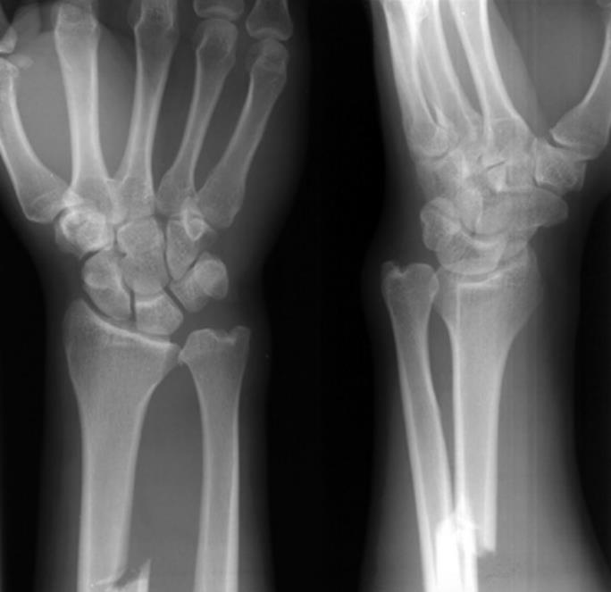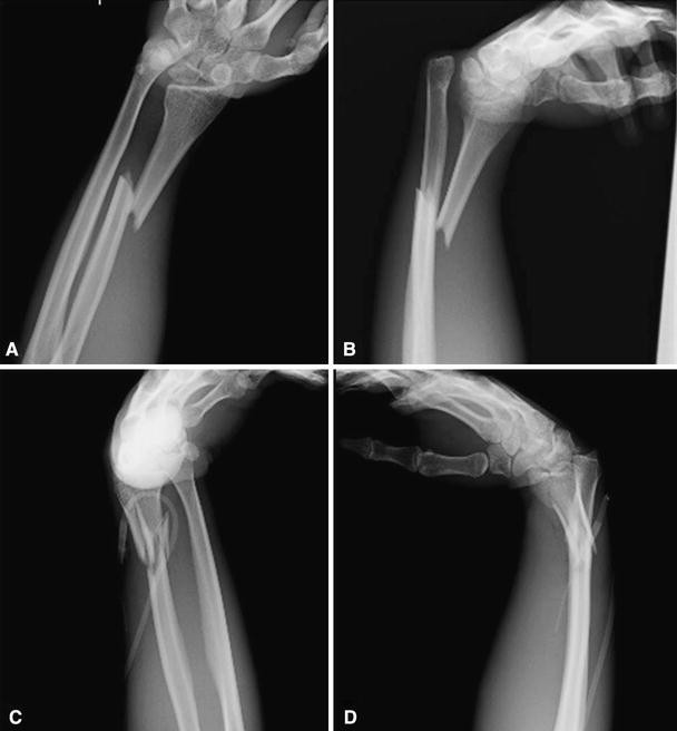
Children bone is softer and more pliable than adults. On the lateral view, the ulnar styloid should point posterior, and the coronoid process should point anterior, whereas the radial styloid and the bicipital tuberosity will not be visible. On an AP view of an uninjured forearm, the radial styloid will be 180° from the bicipital tuberosity with the tuberosity pointed ulnar. New studies evaluate the possibility to use the ultrasound as an accurate method for diagnosis, some advantages could be rapidity, radiation-free and less painful for the patients. Although angulation is easier to measure, rotation can be more difficult. Standard anteroposterior (AP) and lateral orthogonal forearm radiographs are typically sufficient to diagnose a forearm fracture. The elbow and wrist are examined to check for a Monteggia or Galeazzi injury.

Distal pulses and capillary refill are assessed. Abuse should be considered in children younger than 3-years-old. Įxamination of an acute child injured it is not an easy task. The brachioradialis dorsiflexes and deviates radially the distal fragment during a distal third fractures. Fractures that happened in the middle third are altered by the pronator quadratus more distally, which pronates the distal fragment, meantime the impact of bicep on the more proximal fragment is negated by the pronator teres, causing the fragment to remain in neutral position. The supinator and bicep flex and supinate the proximal fragment, when there is a proximal fracture. The bicep attaches proximally at the bicipital tuberosity on the anterior medial radius. Understanding the deforming forces is essential to the reduction in both-bone forearm fractures (Fig. However, single bone forearm fractures of the ulna or radius should always raise suspicion for a Monteggia or Galeazzi fracture dislocation, respectively. Single bone forearm fractures are far less common and are typically the result of direct trauma. Pediatric forearm fractures typically follow indirect trauma, such as a fall on an outstretched hand coupled with a rotational component. Additional remodeling can also be attributed to elevation of the thick osteogenic periosteum after fracture. This polarization of growth shows why distal fractures demonstrate a higher remodeling potential than do fractures closer to the elbow. The distal radial and ulnar growth plates are responsible for 75% and 81% of the longitudinal growth of each bone, respectively.

The interosseous membrane is higher strain proximally in neutral and pronation, and is higher strain distally when in supination. The radial bow, an apex lateral bend in the radius, increases the range of pronation. Radius and ulna are attached by the proximal annular ligament, by the interosseous membrane along the diaphysis, and distally by the ligaments of the distal radioulnar joint and triangular fibrocartilage complex. Anatomically, the ulna is relatively straight and static, it plays a more important role in maintaining forearm stability, especially when subjected to buckling and torsional stress. Understanding pediatric forearm anatomy offers important guidelines for treatment in the nonoperative and operative settings.

Both forearm bones were fractured in 50.1% of cases of forearm injuries and there were significantly more males than females (63.6% vs. found forearm fractures to be significantly more frequent in school age children (65%) and adolescents (63%) compared to infants (42%) and preschool children (50%). Forearm fractures account for 17.8% of all fractures in pediatric age. using the 2010 NEISS report, estimated in children aged 0 to 19 years, 5,333,733 emergency room (ER) visits, of which 788,925 (14.7%) were fracture related. Forearm fractures are the most common type of fractures in the pediatric population, but, to date, no comprehensive overviews of their epidemiology are available.


 0 kommentar(er)
